39 picture of the eye with labels
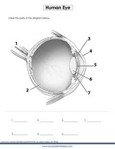
What is an eye mark and why do I need it? - Consolidated Label An 'eye mark' (also known as 'eye spot') is a small rectangular printed area located near the edge of the printed flexible packaging material. A sensor on the form-fill-seal (FFS) machine reads the eye mark to identify packaging material, control the material's position, and coordinate the separation and cutting of the flexible packaging material. Label the Eye - The Biology Corner Label the Eye. Shannan Muskopf December 30, 2019. This worksheet shows an image of the eye with structures numbered. Students practice labeling the eye or teachers can print this to use as an assessment. There are two versions on the google doc and pdf file, one where the word bank is included and another with no word bank for differentiation.
Human Eye Anatomy Pictures, Images and Stock Photos Browse 7,999 human eye anatomy stock photos and images available, or search for vision or retina to find more great stock photos and pictures. Newest results vision retina human eye structure eye chart human eyeball eye doctor eye diagram cataract retinopathy pancreas Anatomy of human eye and descriptions.
Picture of the eye with labels
500+ Stickers for Kids | Oriental Trading Company Hand Eye Coordination Development is Important: Encourage Learning With Stickers For Kids! Help children develop those important skills by incorporating stickers into their learning activities. Use a sticker scene and guide children where to sticker particular stickers, so they can practice hand eye coordination. Eye Anatomy: 16 Parts of the Eye & Their Functions The lens of the eye (or crystalline lens) is the transparent lentil-shaped structure inside your eye. This is the natural lens. It is located behind the iris and to the front of the vitreous humor (vitreous body). The vitreous humor is a clear, colorless, gelatinous mass that fills the gap between the lens and the retina in the eye. Human Eye Coloring Page | crayola.com Human Eye. Use Crayola® crayons, colored pencils, or markers to color the parts of the human eye. Use the word bank below to identify parts of the eye.The eye is the organ that collects images and sends them to the brain, so you can see. The eye is protected by the bones of your skull and six muscles.
Picture of the eye with labels. Label the Eye Worksheet - Teacher-Made Learning Resources - Twinkl The first page is a labelling exercise with two diagrams of the human eye. One is a view from the outside, and the other is a more detailed cross-section. On the second page, you'll find a set of answers showing the properly labelled human eyes, designed to help you check the worksheets without having to come up with your own answer key. Eye anatomy: A closer look at the parts of the eye Eye anatomy: A closer look at the parts of the eye. By Liz Segre. When surveyed about the five senses — sight, hearing, taste, smell and touch — people consistently report that their eyesight is the mode of perception they value (and fear losing) most. Despite this, many people don't have a good understanding of the anatomy of the eye, how ... Eye Diagram - Differentiated Worksheets and EASEL Activities - Pinterest Jan 29, 2016 - Use these simple eye diagrams to help students learn about the human eye. Three differentiated worksheets are included: 1. Write the words using a word bank2. Cut and paste the words3. Write the words without a word bank Labels include: eyebrow, eyelid, eyelashes, pupil, iris, and sclera.UPDATE:I'... The Best Picture Settings for LG 4K TVs - Lifewire Oct 27, 2020 · Eye Comfort Mode: Automatically adjusts the color temperature to reduce eye strain over long viewing periods. HDMI Ultra HD Deep Color: This allows a designated HDMI input to access 4k@60Hz signals encoded with 4:4:4, 4:2:2, or 4:2:0 chroma subsampling. However, if you don't have source devices capable of sending these signals, it's best to ...
Eye Anatomy Diagram - EnchantedLearning.com Definitions : Aqueous humor - the clear, watery fluid inside the eye. It provides nutrients to the eye. Astigmatism - a condition in which the lens is warped, causing images not to focus properly on the retina. Binocular vision - the coordinated use of two eyes which gives the ability to see the world in three dimensions - 3D. Transverse Section Of Eye Anatomy With Labels High-Res Vector Graphic ... Transverse section of eye anatomy with labels. - stock illustration. Transverse section of eye anatomy with labels. Buy the print. Get this image in a variety of framing options at Photos.com. Eye Diagram With Labels and detailed description - BYJUS A brief description of the eye along with a well-labelled diagram is given below for reference. Well-Labelled Diagram of Eye The anterior chamber of the eye is the space between the cornea and the iris and is filled with a lubricating fluid, aqueous humour. The vascular layer of the eye, known as the choroid contains the connective tissue. Eye vs. Camera | Let's Talk Science The photoreceptors in a camera are evenly distributed across the lens. In the human eye, however, the cones are concentrated at the centre of the retina. There are no rods at all at the centre of the retina. How else are your eyes different from a camera? Because a camera has photoreceptors all over its lens, it always sees a “full” picture.
Printable Eye Images | Etsy Vintage image Human Eye Instant Download picture Digital printable clipart graphic Burlap Fabric Transfer Iron On Decor T-shirt 300dpi. Ad by UnoPrint Ad from shop UnoPrint. UnoPrint. From shop UnoPrint. 5 out of 5 stars. (1,869) Sale Price $1.96. $1.96. $2.80. Sparkle: Original Motion Picture Soundtrack - Wikipedia Sparkle: Original Motion Picture Soundtrack is the soundtrack album for the 2012 Sony/TriStar Pictures film Sparkle, a remake of the 1976 film of the same name. The album was released through Sony Music Entertainment 's RCA Records on July 31, 2012. Quiz: Label The Parts Of The Eye - ProProfs Quiz: Label The Parts Of The Eye. People say that the eyes are the windows to a person's soul. In the class today, we covered parts of the eye, and what changes in them should be alarming to a patient. How much did you get to understand about the human eye? The Eye - diagram to label | Teaching Resources File previews. pdf, 2.94 MB. Diagram of eye with key words to use in labelling it. Tes classic free licence.
31 Label The Eye Quiz - Best Labeling Ideas
Eye Diagram Unlabelled - Wiring Diagram Pictures 07.12.2018. 1 Comments. on Eye Diagram Unlabelled. Select the correct label for each part of the eye. The image is taken from above the left eye. Click on the Score button to see how you did. Incorrect answers will. 61 high-quality Unlabeled Eye Diagram for free! human eye diagram. YouTube.

KATMAN SCIENCE
blank eye diagrams - Bing Images | Human eye diagram, Human ear diagram ... Human Eye Diagram Unlabeled. Find this Pin and more on EMS by Melissa Bena. See 12 Best Images of Anatomy Human Ear Diagram Worksheet. Inspiring Anatomy Human Ear Diagram Worksheet worksheet images. Study Digestive System with pictures flashcards.

Fitness and Bodybuilder Model Frank DeFeo | Model Galleries
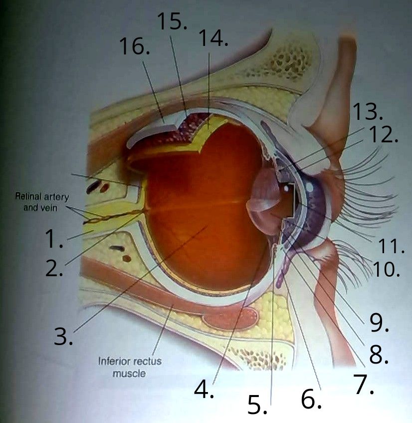
The Human Eye (Eyeball) Diagram, Parts and Pictures The eyeball is a round gelatinous organ that contains the actual optical apparatus. It is approximately 25 mm in diameter and sits snugly in the orbit where six muscles control its movement. The eyeball has three layers, each of which has several important structures that are essential for the sense of vision. Wall of the Eyeball
Label the Part of the Eye -- Exploring Nature Educational Resource
Solved B с A E F D Match the following parts of the eye with - Chegg Science. Anatomy and Physiology. Anatomy and Physiology questions and answers. B с A E F D Match the following parts of the eye with the labels in the picture above. A Iris F Cornea В. Ciliary Muscles G Optic Nerve C Lens E Retina Aqueous and Vitreous Fluid. Question: B с A E F D Match the following parts of the eye with the labels in the ...

30 Label The Eye Diagram - Labels Database 2020
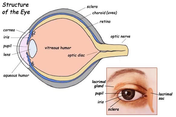
Human Eye Diagram - Human Body Pictures & Images - Science for Kids Photo description: This human eye diagram gives an excellent overview of the human eye. The cross section features labeled parts such as the iris, pupil, cornea, lens, retina, choroid, optic disc, optic nerve and fovea. For more information on eyes, check out our range of interesting human eye facts.

35 Label Of An Eye - Labels Database 2020
Labelling the eye — Science Learning Hub In this interactive, you can label parts of the human eye. Use your mouse or finger to hover over a box to highlight the part to be named. Drag and drop the text labels onto the boxes next to the eye diagram If you want to redo an answer, click on the box and the answer will go back to the top so you can move it to another box.
Eye Flashcards | Easy Notecards
Label Parts of the Human Eye - University of Dayton Parts of the Eye Select the correct label for each part of the eye. The image is taken from above the left eye. Click on the Score button to see how you did. Incorrect answers will be marked in red.

Kate McKinnon Hot Pics and Bio | Picture Perfect
20 Different Ways to Draw the Eye - Improve Drawing Realistic Eye Drawings. Creating a realistic drawing of an eye is a technique that will enable you to break down the eye's form to its basic parts.. This way of drawing the eye is best begun with a faintly drawn line. Draw the eye from direct observation using a mirror or drawing a model.

Eye Colors: Iridology Iris Pictures
Human eye anatomy Images, Stock Photos & Vectors - Shutterstock 60,203 human eye anatomy stock photos, vectors, and illustrations are available royalty-free. See human eye anatomy stock video clips Image type Orientation Sort by Popular Biology Healthcare and Medical Icons and Graphics Recreation/Fitness human eye anatomy 3d rendering eye visual perception infographic Next of 603
Labeling the eye Quiz - Quizizz
PDF Eye Anatomy Handout - National Eye Institute of light entering the eye. Lens: The lens is a clear part of the eye behind the iris that helps to focus light, or an image, on the retina. Macula: The macula is the small, sensitive area of the retina that gives central vision. It is located in the center of the retina. Optic nerve: The optic nerve is the largest sensory nerve of the eye.

My Life in Tupperware Bins: The Life of a Tall Girl
PDF Parts of the Eye - National Eye Institute | National Eye Institute Parts of the Eye . To understand eye problems, it helps to know the different parts that make up the eye and the functions of these parts. Here are descriptions of some of the main parts of the eye: Cornea: The cornea is the clear outer part of the eye's focusing system

Fitness and Bodybuilder Model Frank DeFeo | Model Galleries
Label Eye Printout - EnchantedLearning.com Label the Eye Diagram. Human Anatomy. Read the definitions, then label the eye anatomy diagram below. Cornea - the clear, dome-shaped tissue covering the front of the eye. Iris - the colored part of the eye - it controls the amount of light that enters the eye by changing the size of the pupil. Lens - a crystalline structure located just behind ...
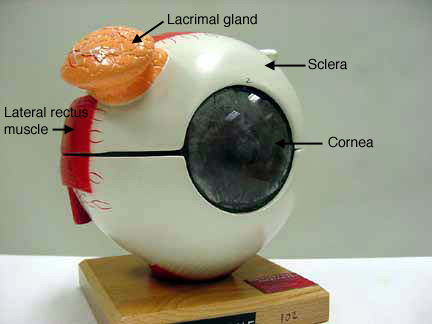
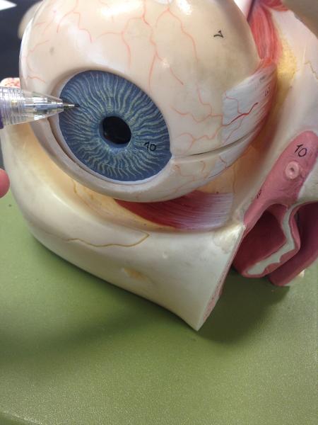
Apparently not even the professor knows what this part of the eye is. The tan lump in this model ...
Eye Anatomy Detail Picture Image on MedicineNet.com Picture of Eye Anatomy Detail The eye is our organ of sight. The eye has a number of components which include but are not limited to the cornea, iris, pupil, lens, retina, macula, optic nerve, choroid and vitreous. Cornea: clear front window of the eye that transmits and focuses light into the eye.
Realistic butterflies vectors
A Picture of the Eye - WebMD One eye sees better than the other, so your brain favors that eye. The weaker eye, which may or may not wander, is called the "lazy eye." Astigmatism : A problem with the curve of your cornea.

The BioLogs: CSEC - The Eye - functions of the various parts
CUT-AND-ASSEMBLE PAPER EYE MODEL • thin permanent marker for a number labels on plastic parts (such as a very thin point Sharpie) Assembly: 1) After copying pattern pages onto card stock, cut out all parts. On the background page that says THE HUMAN EYE, cut away the black rectangles and trim the triangles at the bottom, as shown in picture above.

Quiz – Page 2
Structure and Functions of Human Eye with labelled Diagram Human Eye Diagram: Contrary to popular belief, the eyes are not perfectly spherical; instead, it is made up of two separate segments fused together. Explore: Facts About The Eye To understand more in detail about our eye and how our eye functions, we need to look into the structure of the human eye.

Jens Öhman by Franz Fleissner | Oh yes I am
Human Eye Coloring Page | crayola.com Human Eye. Use Crayola® crayons, colored pencils, or markers to color the parts of the human eye. Use the word bank below to identify parts of the eye.The eye is the organ that collects images and sends them to the brain, so you can see. The eye is protected by the bones of your skull and six muscles.

World's Beautiful things around us !: Beautiful nature| Eye cooling pictures every body loves to ...
Eye Anatomy: 16 Parts of the Eye & Their Functions The lens of the eye (or crystalline lens) is the transparent lentil-shaped structure inside your eye. This is the natural lens. It is located behind the iris and to the front of the vitreous humor (vitreous body). The vitreous humor is a clear, colorless, gelatinous mass that fills the gap between the lens and the retina in the eye.
Post a Comment for "39 picture of the eye with labels"